Skip to content

Research

Translational Research Model

Our laboratory studies the skin, which serves as a protective shield between the human body and its environment. This critical barrier tissue is tasked with sealing the body to prevent water loss, excluding pathogens like bacteria to avoid infection, and withstanding damage from environmental insults like ultraviolet radiation to resist cancer formation. We focus on the biology of skin cells called keratinocytes with the goal of understanding how they normally mature to form an effective barrier tissue and how they are compromised in skin disease. Our ultimate goal is to identify novel approaches to promote skin tissue regeneration, to prevent skin damage from environmental stressors, and to treat genetic dermatologic disorders.

Keratinocyte Cell Biology

The outermost layer of the skin, the epidermis, is made up of multiple layers of cells called keratinocytes that form a protective barrier for the body. Keratinocytes continually replicate themselves to replenish the skin tissue as it is naturally shed every month. While keratinocytes slowly move outward toward the skin surface, they must undergo a unique process of maturation to harden and strongly adhere to one another to protect the body and resist wounding. Unfortunately, keratinocyte maturation is disrupted in many human skin diseases like eczema and psoriasis or in rare genetic disorders like ichthyoses. Our limited understanding of the fundamental biology of keratinocyte maturation has prevented development of effective therapies to promote regeneration of the epidermis after wounding or for patients with genetic skin disorders.

Epidermis Diagram

Organoid Skin Imaging

Our lab has developed an “organoid” model of the skin in which human keratinocytes form a multi-layered epidermal tissue in just one week. Coupling this system with high-resolution imaging techniques, like confocal microscopy, permits us to visualize changes in the live organoid skin model as it is developing into a mature tissue. We can engineer human keratinocytes to express fluorescent proteins that are visible using a high-magnification microscope allowing us to visualize these cells at the level of single organelles (e.g., mitochondria) to better understand how the epidermis forms.

Skin Organoid Model
Skin Organoid Imaging

Gene Editing in the Skin

We use gene editing techniques (e.g. CRISPR/Cas9) in human keratinocytes to introduce DNA mutations linked to dermatologic disorders. By culturing these engineered cells in the organoid system, we are able to replicate skin tissue pathology in the lab. Such 3-D models of skin disease can then be used for high-resolution imaging to better understand the underlying pathophysiology and for testing new treatment approaches. Our ultimate goal is to identify novel therapies to promote tissue regeneration after skin injuries and to restore epidermal barrier function in inherited and currently incurable dermatologic diseases.

Gene Editing in Skin

Featured News

Congratulations Jessica Ayers & Crystal Tam

Please join us in congratulating Simpson Lab student researchers, Jessica Ayers and Crystal Tam! Jessica, a M3D PhD…

Read more

Dr. Cory Simpson Receives ASCI Young Physician-Scientist Award

Originally published to the UW Department of Dermatology website on Jan. 26, 2024. UW Dermatology is proud to…

Read more

About the Simpson Lab

Dr. Simpson launched a research laboratory at the University of Washington in 2021 to study epidermal differentiation and how it is compromised in dermatologic disease. The Simpson Lab focuses on the role of autophagy and organelle degradation in human keratinocytes and how these cellular pathways drive maturation and homeostasis of the epidermal tissue. Using an organotypic model of human skin coupled with advanced microscopy, his lab can visualize the live tissue at the level of single organelles to better understand how the epidermis forms and how disease-associated mutations disrupt the tissue. Ultimately, the Simpson Lab aims to identify novel treatment strategies to promote epidermal tissue regeneration after skin injuries and to restore skin barrier function in inherited and currently incurable dermatologic diseases.

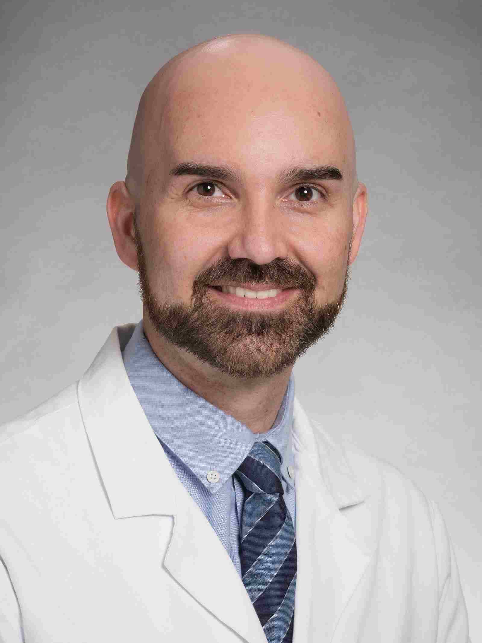
The Simpson Lab is located at the UW Medicine South Lake Union (SLU) Campus amidst an innovative biomedical research hub in Seattle including the Allen Institute for Cell Science just across the street. The SLU Campus is connected to the Fred Hutchinson Cancer Research Center and the main UW campus by a convenient shuttle. Dr. Simpson is a member of the Institute for Stem Cell and Regenerative Medicine (ISCRM) at UW, which offers access to outstanding core resources for imaging, stem cells, genomics, and high-throughput screening.

Dr. Simpson’s research has been supported by the National Institutes of Health (NIH), the National Institute of Arthritis and Musculoskeletal and Skin Diseases (NIAMS), the Dermatology Foundation, the American Skin Association, the National Psoriasis Foundation and the La Roche-Posay North American Foundation.

Back To Top