
New Simpson Lab paper in JCI Insight links epidermal autophagy to Darier disease
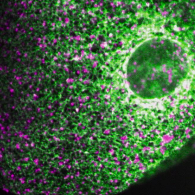
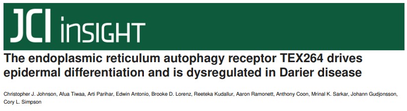
The Simpson Lab published a new paper in JCI Insight that utilized live microscopic imaging of a 3D human skin model to understand how keratinocytes mature to form the epidermal barrier and how this process is impaired in Darier disease, a rare epidermal differentiation disorder that Dr. Simpson sees in his dermatology clinic.
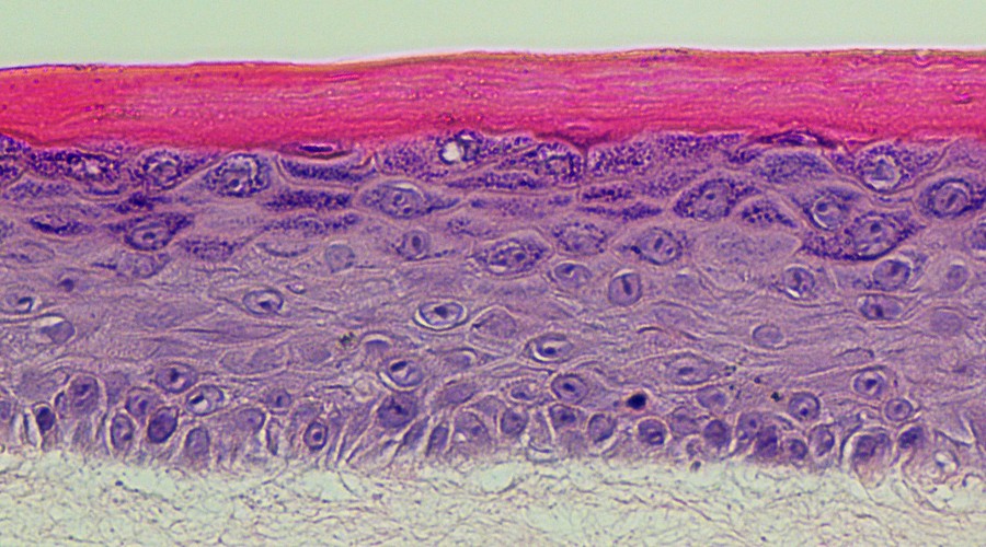
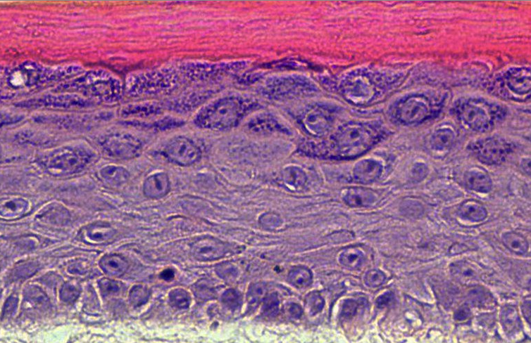
The epidermis is made of multiple layers of keratinocytes that progressively mature as they move outward and eventually die to form the dry, flaky layers that shed from the skin. Toward the end of their life, keratinocytes degrade their nuclei and organelles, which allows them to flatten into the compacted outer layers of dead cells (corneocytes) that seal the body off from the environment.
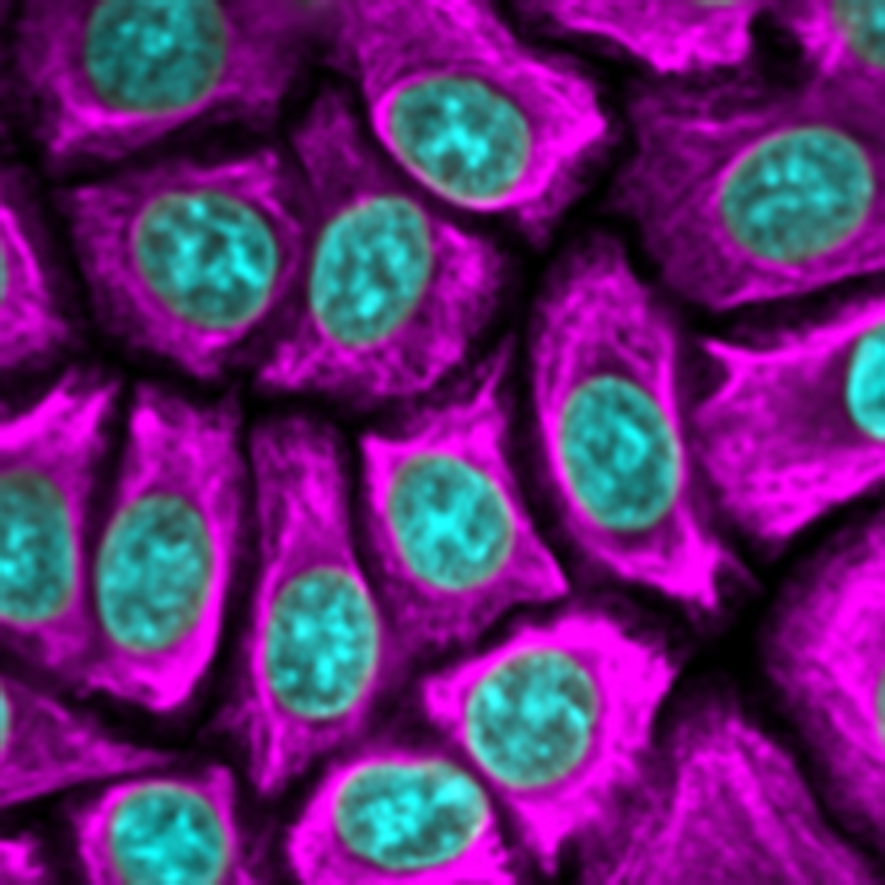
The Simpson Lab's project zoomed in on the endoplasmic reticulum (ER), applying a confocal microscope to their human epidermal tissue model to reveal how keratinocytes break down this massive network of tubules. Dr. Simpson's team found that keratinocytes activate a specialized cellular degradation pathway called autophagy to get rid of the ER. Upon reaching the outermost layers of the epidermis, keratinocytes turned on an autophagy protein called TEX264, which cut the ER tubules into bits and put them into the cell's garbage disposal, the lysosomes.
Connecting these basic science findings to his clinical expertise, Dr. Simpson showed that dysregulation of the autophagy pathway may explain the abnormal epidermal maturation (dyskeratosis) seen in biopsies of Darier disease, which causes painful skin blistering and infections in patients. Whether manipulating autophagy with existing drugs could be therapeutic in Darier disease is currently being tested.
The project was led by UW School of Medicine alumnus Christopher (CJ) Johnson, M.D., who matched in a research-track dermatology residency at the University of Colorado after completing a gap-year in the Simpson Lab funded by an ASCI Physician-Scientist Support Fellowship. The work was also supported by grants from the National Institutes of Health, the American Skin Association, the Foundation for Ichthyosis and Related Skin Types, the LEO Foundation, the National Psoriasis Foundation, and the UW Institute for Stem Cell and Regenerative Medicine.